The mechanism of formation of vestibulo-cerebello-ophthalmic reactions and vestibulo-cerebellospinal reactions in rotation tests. A new theory of kinetic stimulations.
In 1873 three scientists working independently: Ernst Mach; Josef Robert Breuer and Crum Brown presented the hydrodynamic theory which maintains that the head movement causes the flow of endolymph in semi-circular canals of the labyrinth and the sensual elements in the canals are stimulated by the fluid movement of changes of pressure (1). Within the ampullae of the semi-circular canals there are receptor organs built from ampullar crests, supplied with ciliary cells reacting to the endolymph movement. The ciliary epithelium includes support cells and ciliary cells. The ciliary cell comprises several dozen thin cilia of different length called stereocilia and one cilium, situated on the border, thicker and the longest called kinocylium. The fact that the receptor organ (the cupula) is located in the endolymphatic fluid means that its kinetics while using accelerations or delays should be treated as the pendular movement in the liquid environment (2). It is known that at rest ciliary cells in horizontal canals possess the so-called resting potential whose impulsation is about 5-25 cycles/s. This impulsation is transmitted to the central equilibrium system and maintains the constant, symmetrical muscular tonus - fig. 1.
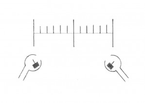
Fig. 1. The diagram of the resting activity of both labyrinths.
Both horizontal canals behave antagonistically, in the restful conditions they remain in the equilibrium and therefore eyeballs remain in the indirect position. The most effective flow of intralabyrinthine fluids in semi-circular canals is obtained when the plane of a suitable semi-circular canal is compatible with the plane of rotation. Such an optimum for the horizontal canals is obtained when the subject is sitting in the chair with his/her head bent by about 30° to the chest. As known from the history of discoveries in the field of otoneurology, Ernst Ewald summed up his own wide experimental research on animals in two laws. The second Ewald's law says that in semi-circular horizontal canals the ampulopetal flow of the endolymph causes a considerably stronger nystagmus reaction than the ampulofugal (intraspinal) flow. Inverse reactions occur while irritating the vertical canals. For many years this law has been universally accepted by labyrinthologists and it was interpreted that the condition of causing a reaction from the semi-circular horizontal canal is caused by the ampulopetal flow of endolymph. Such an interpretation suggested that the ampullar organ was a receptor of powers acting only in one direction. LeDoux (3) put forward a conclusion that the ampulofugal flow of endolymph also causes a reaction which consists in reducing the value of the resting potential of the canal irritated in this manner. Such a situation reveals the resting potential of the horizontal contralateral canal which, as not being irritated in the test directly, obtains in this way the bioelectric predominance. Such an explanation of bioelectric changes in both horizontal canals allows explaining why, during angular accelerations, there appears the phenomenon of nystagmus although the movement of endolymph in one canal occurs towards the utricle, and in the other - towards the canal. The element primarily responsible for the nystagmus reaction is one of the canals towards which there is a rotation with the determined angular acceleration (3, 4). According to this line of thought it becomes clear that during rotation to the right with the determined angular acceleration, the emerging moment of inertia of liquid will cause the movement of endolymph towards the opposite, it means to the left, thereby causing inclination of the cupula on the right side towards the utricle (an increase of potential) - Fig. 2.
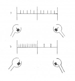
Fig. 2. The diagram of the activity of the labyrinths: a) - at rest. b) - during rotations to the right according to the theory by LeDoux, Montandon and Russbach. The visible increase of the activity of the right labyrinth and the decrease of the activity of the left labyrinth.
Therefore, it has been maintained by now that the bioelectric predominance in the physiological conditions always arises on the side compatible with the direction of the acceleration, causing rotatory nystagmus directed to the same side. Knowing the previous theories explaining the formation of nystagmus reactions in kinetic stimulations, the question is how one should explain the obtainment of symmetrical reactions after the unilateral entire destruction of the labyrinth in the period of compensation of post-rotary reactions of nystagmus after rotations to the left and right. With identical constant stimulations during rotations to the right and left, the same responses are received, although the only irritated labirynth is the healthy one, in which the endolymph flow keeps the ampulopetal and ampulofugal direction. Based on the previous knowledge of such a logical explanation cannot be introduced. Impulses coming from the cupula organs as a result of kinetic stimulation to vestibular nuclei cause motor reactions of the reflexive type. At that time the vestibulo-ophthalmic reflex and the vestibulospinal reflex appear completely independent from our will. Robert Barany (1) mentions that earlier, before he had begun his research it was considered that reflexes did not occur directly but through the cerebellum. Therefore these reflexes must be called the vestibulo-cerebello-ophthalmic reflexes and the vestibulocerebellospinal reflex. By now nobody has presented any description of such reflexes. Barany (1) proved that the direction of the deviation of limbs always occurs contrary to the appearing nystagmus. The deviation reaction always concerns all of the four limbs. In this way he succeeded in proving that the direction of the movement is located in the cerebellum. In his collection he found cases in which the reaction of the limbs did not appear only towards the damaged cerebellar hemisphere. The researcher found that nerve cells and nerve fibers connected with a certain direction must lie near one another in the cerebellar hemisphere. On the basis of the gathered experiments Barany arrived at the final conception of the principles of operation of the cerebellum. He assumed that in the cerebellum there are four centers connected with the muscular apparatus, as regard the direction of the movement - one center to the right, one to the left, one upwards and one downwards. In healthy people, as long as there is no stimulation of the horizontal canals, these four centers, which have certain tension called the tonus, balance one another and neither nystagmus nor the deviation of limbs is observed. The earlier Nakiela's research (5) concerning a comparative assessment of nystagmus reactions obtained in the rotation test, according to Arslan in healthy people and in ill people with unilateral canal paralysis in the period of obtaining symmetrical nystagmus reactions to the right and left showed that the average values of individual parameters of the post-rotary nystagmus in the group of ll persons were by half smaller than in the control group of healthy persons. For example, the average duration of the post-rotary reaction in healthy people was 34 seconds, the average quantity of saccadic eye movement was 66. In the group of ill people the average duration of the post-rotary reaction was 17 seconds, the average quantity of saccadic eye movement was 33. Then the author drew a conclusion that both labyrinths are equally responsible for the development of the post-rotary reaction in healthy people. This undermined the theory by LeDoux, Montandon and Russbach (3, 4), which is still binding in otoneurology. This is logical, as the energy of one labyrinth was destructed then the received reactions must be by half smaller. I will present the explanation of the mechanisms of formation of vestibulo-cerebello-ophthalmic reflexes and vestibulo-cerebellospinal reflexes based on Figure No.9 which was shown in the habilitation lecture (6).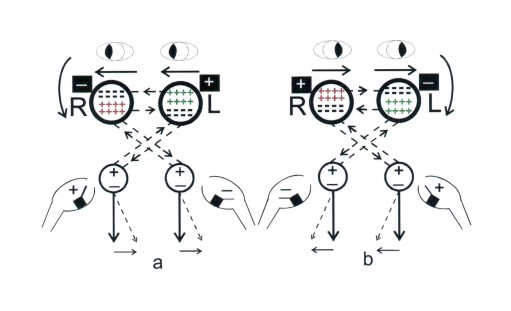
Fig. 9 Stimulation of labyrinths in the rotation test. The diagram of formation of the vestibulo-cerebello-ophthalmic reactions and vestibulo-cerebellospinal reactions during the rotations: a) to the right b) to the left
During the rotations to the right with a suitable angular acceleration, in the right horizontal canal of the labyrinth there is an endolymph flow towards the ampulla, which causes kinocilium to deviate towards the utricle. There is an increase in positive bioelectric potential which is transmitted to the right complex of vestibular nuclei and then to the left cerebellar hemisphere. The left cerebellar hemisphere is stimulated, which is symbolically marked with the "plus" sign. From the stimulated left cerebellar hemisphere impulses are transmitted back to the complex of vestibular nuclei on the right side. From this complex of vestibular nuclei the bioelectric potential is transmitted via the ascending tract by the medial long bundle to nuclei of motor cranial nerves III, IV and VI, supporting the external muscles of eyeballs. This situation leads to the right abducent nerve being stimulated, thanks to which the right eyeball is taken away outside and nystagmus appears to the right with a suitable power vector directed to the right. In this way there appears a vestibulo-cerebello-ophthalmic reflex. Simultaneously, from the right complex of vestibular nuclei, the bioelectric potential is transmitted to the spinal cord by means of the vestibulospinal lateral tract and the vestibulospinal medial tract. Then the muscles of adductors of the upper limb and the lower are stimulated. During the examination we can observe adducting of the right upper limb. As a result a vestibulo-cerebellospinal reflex appears. At the same time in the left horizontal canal of the labyrinth there appears the endolymph movement from the ampoule, which results in kinocilium leaning towards the canal. There appears a growth of the negative bioelectric potential which is transmitted to the left complex of vestibular nuclei and then to the right cerebellar hemisphere. The right cerebellar hemisphere is inhibited, which was symbolically marked with the "minus" sign. From the right cerebellar hemisphere impulses are returned to the vestibular nuclei complex on the left side. From this vestibular nuclei complex the bioelectric potential is transmitted via the ascending tract by the medial long bundle to the nuclei of motor cranial nerves III, IV and VI, supporting the external muscles of eyeballs. In this situation the left oculomotor nerve is stimulated, thanks to which the left eyeball is adducted and there appears nystagmus to the right with identical power vector directed to the right as on the right side. Consequently, a vestibulo-cerebello-ophthalmic reflex appears on the other side. This example helps us understand that both the right and the left labyrinth are equally responsible for nystagmus to the right. Simultaneously, from the left complex of vestibular nuclei the bioelectric potential is transmitted to the spinal cord by means of the lateral vestibulospinal track and the medial vestibulospinal track. The left upper limb and the lower limb is abducted. During the research we observe the abduction of the left limb to the outside. This results in a vestibulo-cerebellospinal reflex from the left labyrinth. During the rotations to the right with a suitable angular acceleration there appears rotatory nystagmus to the right and a deviation of upper limbs to the left. Figure 1 presents the diagram of the activity of labyrinths according to Nakiela.
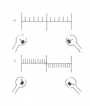
Fig. 3 The diagram of the activity of labyrinths according to Nakiela: a) at rest b) during rotations to the right - a visible increase in positive potentials in the right labyrinth and an increase in negative potentials in the left labyrinth.
Vestibular nuclei and the cerebellar hemispheres are a closed system of feedbacks. Vestibulo-cerebello-ophthalmic reflexes and vestibulo-cerebellospinal reflexes will be realized inversely during rotations with a suitable angular acceleration to the left (fig. 9b).
As I mentioned earlier, a vestibulo-cerebello-ophthalmic reflex and a vestibulo-cerebellospinal reflex is an automatic reflex, completely independent from our will. The main central organ of the equilibrium system is the cerebellum. How are then the vestibulo-cerebello-ophthalmic reflexes and vestibulo-cerebellospinal reflexes shaped during rotations to the right with a suitable angular acceleration in the event of the complete dematerialization of the right labyrinth. These mechanisms will be explained based on fig. 10 presented in the habilitation lecture.
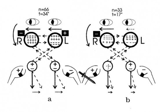
Fig. 10
Fig. 10 Stimulation of labyrinths in the rotation test. The diagram of formation of the vestibulo-cerebello-ophthalmic reaction and vestibulo-cerebellospinal reaction during rotations to the right:
a) in a healthy person
b) in an ill person after destruction of the right labyrinth in the period of compensation himself nystagmus reactions at rotations to the right and left.
n - the number of saccadic eye movements during the post-rotary reaction
t - the duration of the post-rotary reaction
The diagram of formation of the vestibulo-cerebello-ophthalmic reactions and the vestibulo-cerebellospinal reactions presented in fig. 10a is identical as the one resented in fig. 9a. In conclusion, both labyrinths are equally responsible for the development of the nystagmus reaction during rotations to the right.
During rotations to the right with a suitable angular acceleration in an ill person with a complete destruction of the right labyrinth (fig. 10b) we deal with a different situation.
As it is known, during the destruction of the right labyrinth there appears a paralytic nystagmus to the left, i.e. towards the healthy labyrinth. In this case, in the early phase of paralysis during rotations to the right and left we receive only a nystagmus reaction to the left. As soon as the right labyrinth is damaged, the process of habituation begins which will be explained in detail further in the lecture. The constant resting impulsation coming from the left labyrinth causes constant stimulation of the right cerebellar hemisphere and causes the paralytic nystagmus to the left. The body defends itself against a superfluous loss of energy for creating the paralytic nystagmus and therefore from the beginning of the injury the process of habituation is triggered. The constant stimulation of the right cerebellar hemisphere through restting stimuli coming from the left labyrinth causes the right cerebellar hemisphere to become inhibited, that is in the right cerebellar hemisphere an increased number of negative signs begins to appear and in the left hemisphere simultaneously - an increased number of positive signs. It creates a new situation when the right cerebellar hemisphere is inhibited and the left one is stimulated. The resting stimulations from the left labyrinth go to the inhibited right cerebellar hemisphere and cause an increasingly weaker reaction, spontaneous nystagmus to the left gradually disappears, a spontaneous nystagmus towards the damaged labyrinth often begins to appear, which means that the directional predominance to the right is developed. In rotation tests, in a suitable period, we can obtain symmetrical nystagmus reactions in rotations to the right and left. If the process of habituation is very strongly developed, then in post-rotation tests we can obtain only nystagmus reactions directed toward the damaged labyrinth, in a situation when the endolymph movement in the healthy labyrinth occurs ampulofugally. When the endolymph movement in the healthy labyrinth occurs ampulopetally we may receive no post-rotary reaction. As introduced in fig. 10b, during rotation to the right with a specified angular acceleration, there appears nystagmus to the right and a deviation of upper limbs to the left. During rotations to the right, in the semi-circular horizontal canal there is an endolymph flow in the ampulopetal direction and kinocillium is bent towards the utricle. Since the right labyrinth was completely damaged, then no bioelectric impulse will be transmitted to the nuclear complex on the right and then to the left cerebellar hemisphere. Because, as I presented it before, the left cerebellar hemisphere is in the state stimulation in connection with the phenomenon of habituation, then the bioelectric stimuli from this hemisphere are transmitted to the vestibular nuclei complex on the right, maintaining them in the state of stimulation. Next, as I presented in fig. 9a, reflexes vestibulo-cerebello-ophthalmic and vestibulo-cerebellospinal will be started via the ascending and the descending tract. It will result in nystagmus to the right and the adduction of the right upper limb. These are spontaneous reflexes connected with the phenomenon of habituation and not a result of the reflex caused by the induced rotary stimulus. On the left there will appear an endolymph flow towards the canal and an inclination of the kinocillium also towards the left semi-circular horizontal canal. There appears a bioelectric negative impulse, which is then transmitted to the vestibular nuclei complex on the left and then to the right cerebellar hemisphere causing its inhibition. Because the right cerebellar hemisphere is in the inhibition position, contains symbolically a lot of negative signs, then the stimulus from the left labyrinth is largely strengthened. After that the strengthened bioelectric stimuli from the right cerebellar hemisphere flow to the vestibular nuclei complex on the left. From the complex they are transmitted via the ascending tract by the medial longitudinal bundle to the motor nuclei of the cranial nerves III, IV and VI supporting the external muscles of the eyeballs. Nystagmus to the right is caused. Simultaneously from the left vestibular nuclei complex, the bioelectric potential is transmitted to the spinal cord by means of the lateral and medial vestibulospinal tract. The left upper limb is abductions to the outside. In this manner there appear vestibulo-cerebello-ophthalmic reflexes and vestibulo-cerebellospinal reflexes at an ill person with the destruction of the right labyrinth within a period of the performed habituation. Therefore the spontaneous reaction which arose as a result of habituation from the right nuclear complex and the reaction induced by a rotary stimulus from the left nuclear complex will be compatible. The vectors of these powers act in the same direction. Because the energy of the right labyrinth was completely damaged, the post-rotary reactions after rotation to the right and left will be by half smaller than in a healthy person. As indicated, the quantity of inclinations of eyeballs and the duration of the reaction in post-rotation tests is by half smaller. At the complete destruction of the right labyrinth and rotations to the left with a specified angular acceleration we will have a different situation - fig. 10c..
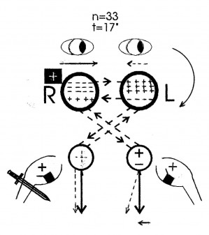
Fig. 10c
Fig. 10c The diagram of formation of the vestibulo-cerebello-ophthalmic reaction and vestibulo-cerebellospinal reaction during rotations to the left in an ill person after the destruction of the right labyrinth in the period of the performed habituation (symmetrical nystagmus reactions in post-rotation tests).
In the period of the performed habituation, so, when obtaining symmetrical post-rotary reactions to the right and to the left, during rotations to the left the endolymph movement in the right semi-circular canal will cause an inclination of the kinocillium towards the canal. In this case, no bioelectric stimulus will flow from the right semi-circular canal to the right vestibular nuclei complex because the labyrinth was completely damaged. Since as a result of the performed habituation the left cerebellar hemisphere was considerably stimulated, the stimuli constantly come from the stimulated left cerebellar hemisphere to the right of vestibular nuclei complex, maintaining in them a constant positive voltage (the plus sign was marked with a dashed line). These nuclei are systematically stimulated and stimuli from them are transmitted via the ascending and the descending tract, as it was shown in figure 10b during rotations to the right. On the left, during rotation to the left with a specified angular acceleration there appears an endolymph movement towards the ampulla, which causes kinocillium to bend towards the utricle. It is followed by an increase in positive bioelectric impulses which are transmitted to the vestibular nuclei complex on the left and then to the right cerebellar hemisphere. Since the right cerebellar hemisphere is in a state of increased inhibition, bioelectric stimuli stimulate it to a lesser degree than normally. Weakened stimuli go back to the vestibular nuclei complex on the left and then via the ascending tract across the medial longitudinal bundle get to the nuclei of the motor cranial nerves III, IV and VI, supporting the external ocular muscles. Next nystagmus directed to the left is caused. Simultaneously, from the left vestibular nuclei complex the positive bioelectric potential is transmitted to the spinal cord via the lateral and medial vestibulospinal tract. The left upper limb is adducted. Since the spontaneous reflexes from the right nuclear complex cause adduction of the right upper limb and nystagmus to the right and reflexes triggered during the stimulation of the left semi-circular canal cause nystagmus to the left and the adduction of the left upper limb, therefore these reflexes are opposed, vectors of these powers act in opposite directions. Finally, in this reaction there will appear nystagmus to the left and the adduction of the left upper limb. The reflex caused by an induced rotary stimulus from the left labyrinth is stronger from the spontaneous reflex from the right vestibular complex and therefore the right limb will remain still. The abduction of the right upper limb right is prevented by the left cerebellar hemisphere stimulated as a result of habituation. Thus, during rotations to the right we obtain nystagmus to the right and a deviation of both upper limbs to the left. During rotations to the left we obtain rotatory nystagmus to the left and only the left upper limb is adducted. When the right labyrinth is damaged within the period of the performed habituation, stimuli coming from the ampulopetal stimulation of the left labyrinth are inhibited, however, stimulus coming from the ampulofugal stimulation of the left labyrinth is strengthened. To sum up, every horizontal canal of the labyrinth is coupled with the cerebellar hemisphere of the opposite side. In the course of kinetic stimulations one of the horizontal canals, in which the endolymph movement occurs towards the utricle, is responsible for the stimulation of one cerebellar hemisphere, while the other, in which the endolymph movement occurs towards the canal, is responsible for the inhibition of the second cerebellar hemisphere. The stimulation of the cerebellar hemisphere in healthy persons always triggers nystagmus towards the opposite hemisphere, the inhibition of the cerebellar hemisphere causes nystagmus towards the stimulated hemisphere. The equal vectors of these powers act in the same direction. The rotatory nystagmus reaction and the post-rotary nystagmus reaction to the right and left in healthy persons is equally caused by the right and the left labyrinth. In ill persons with the unilateral entire destruction of the labyrinth, the nystagmus reaction induced by a rotatory stimulus and the post-rotary stimulus to the right and left is caused only by the unimpaired labyrinth. The size of the nystagmus reaction is then smaller by half than reactions obtained with correctly functioning both labyrinths. The nystagmus reaction always arises only as a result of stimulation of one cerebellar hemisphere, i.e the one which is connected with the healthy labyrinth. At the entire destruction of the labyrinth within the period of the performed habituation, i.e. when in post-rotation tests symmetrical we are obtain nystagmus reactions, during rotations towards the damaged labyrinth with a specified angular acceleration we obtain nystagmus towards the damaged labyrinth and a deviation of both upper limbs towards the healthy labyrinth. However, a rotation towards the healthy labyrinth with a specified angular acceleration will cause a nystagmus reaction towards the healthy labyrinth and adduction of the upper limb on the side of the healthy labyrinth. The limb on the side of the damaged labyrinth remains still. According to the presented principles of operation of the vestibulocerebellar system, the Ewald's 2nd law and the theory of kinetic stimulations according to LeDoux, Montandon and Russbach seem to be false.
References:
1. Barany R. Some new methods for functional testing of the vestibular apparatus and the cerebellum. Nobel Lecture, September 11, 1916.
2. Latkowski B. Rotary tests and their diagnostic meaning. In: Clinical otoneurology. ed. Janczewski Grzegorz. PZWL 1986.
3. LeDoux A. Les canaux semi-circulaires. Acta Oto-Rhino-Laryngol. Belg. 1958, 12, 119-336.
4. Montandon A., Russbach A.: Etude de Quelques syndromes vestibulaires par L'epruve giratoire liminaire. Acta Otolaryngol.(Stockh.). 1956, 46, 264-276.
5. Nakiela J.: Study of the usefulness of the modified Unterberger test in determining efficiency of the equilibrium system. Doctoral thesis. ŁódĽ, Military Medical Academy, 1978.
6 Nakiela J.: The vestibulo-cerebellar system according to the latest investigations and interpretation of the author. Lecture at the examination for the degree of assistant professor. ŁódĽ, Military Medical Academy. 1990.